Albanien
 Bosnien-Herzegowina
 Bulgarien
 Kroatien
 Zypern
 Montenegro
 Tschechien
 Dänemark
 Estland
 Spanien
 Kosovo
 Litauen
 Lettland
 Luxemburg
 Mazedonien
 Deutschland
 Polen
Polen
 Portugal
 Rumänien
 Serbien
 Slowakei
 Slowenien
 Schweiz
 Schweden
 Türkei
 Ukraine
 Ungarn
 Großbritannien
 Italien
 UAE
UAE
 Hong Kong
 Ägypten


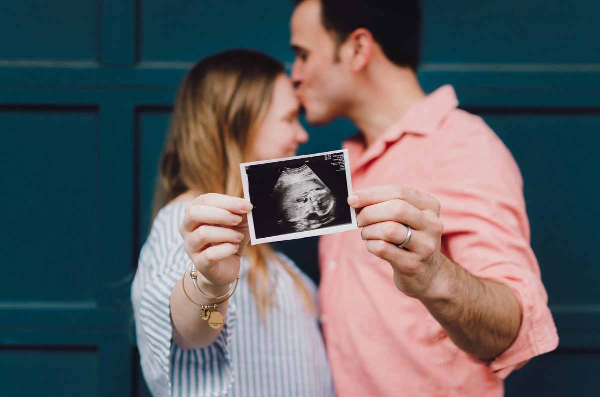
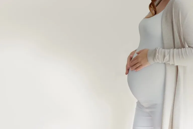
In recent years, the 3D ultrasound has become increasingly popular among parents-to-be. It is undoubtedly a great keepsake for many years to come, but the question is whether it can replace the classic ultrasound in pregnancy? Find out whether a 3D ultrasound in pregnancy is worthwhile and when is the best time for such an examination.
3D ultrasound in pregnancy – what is it?
3D ultrasound is a three-dimensional ultrasound examination that is increasingly used in gynaecological practices. This examination is performed with the same device and in the same way as the classic 2D ultrasound. However, a three-dimensional image is obtained, i.e. its length, width and depth are assessed. Based on the measurements taken, the camera gives the appearance of the structure in question and the doctor (and parents) can have a close look at it.
3D ultrasound in pregnancy – when should it be done?
The best time to have a 3D ultrasound scan during pregnancy is around the 20th week, which is about halfway through the pregnancy. At this time it is easiest to image the baby. The more advanced the pregnancy and the bigger the baby is, the more difficult it is to get a good image. Technical difficulties such as an awkward position of the baby in the uterine cavity or excess fat on the expectant mother should also be taken into account. The volume of the amniotic fluid also affects the quality of the image.

3D ultrasound in pregnancy – is it worth it?
For most parents, the 3D ultrasound is a chance to see the baby “realistically” while it is still in the uterine cavity. They often can’t see much on a classic ultrasound image. 3D ultrasound is also a great souvenir, especially if the doctor captures a smile or a grimace on the baby’s face.
According to experts, 3D ultrasound should not replace grey cell ultrasound, but is an excellent complement to the classic examination. It is particularly useful when a child is suspected of having a malformation such as a cleft lip and palate or polydactyly (abnormal number of fingers). The 3D ultrasound enables the specialist to detect anatomical anomalies in detail because the image is more accurate than with the classic ultrasound.